 From the Desk of Gus Mueller

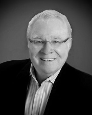
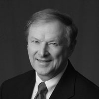
When we think of the study of auditory processing as it relates to neuroanatomy and neuroscience, audiologists generally fall into one of two groups: those who immerse themselves in the laboratory study of the brain, often with animal work, and those who have more of a clinical slant and study it from a functional standpoint. Rarely, do we find an audiologist whose career has excelled in both areas. We are fortunate to have such a person with us this month—internationally known expert Frank Musiek, PhD. Not only can he perfectly dissect the temporal lobe blindfolded (so I’ve heard), he has developed several clinical tests of central auditory function which are commonly used today.
Dr. Musiek is Professor, Department of Speech, Language and Hearing Sciences, University of Arizona. He has a long list of awards, but two that are especially notable are the ASHA Honors of the association, and the AAA Jerger Career Award for research in audiology. We all are familiar with his many popular books on the topics of hearing science, anatomy and physiology, and central auditory processing.
Some of you probably remember Frank from his days at Dartmouth, for others, it might be his many years at UCONN. I recall visiting him at UCONN in the winter of 2014 when he told me he was retiring and going somewhere warm! That decision did indeed lead to warmth, but it also added another 7 years to his career at the University of Arizona. He again is telling me that he is retiring, so we’ll see how it goes this time. I asked him to reminisce a little regarding his career:
Looking back, there were some specific things that were important to me throughout my career. One was combining clinical and basic science in my academic and clinical endeavors. Doing animal and human work provides a perspective on hearing and hearing disorders that is difficult to appreciate unless one has experienced it. I wish all students could gain that kind of experience. It seems that the gulf between clinical and basic science has become greater, which is not what I like to see. I have tried to be a bridge between the basic and clinical sciences; to carry the import of the clinician to the basic scientist and vice versa.
An important part of my career was that I had the opportunity to see neurologically involved patients, both clinically and as research participants. It has been a great experience and I learned a lot. I wish more audiologists could see these fascinating patients. I have tried to champion the clinical audiologist. The clinician is the face of our profession. They need to be supported and their work highlighted. -Frank Musiek
Frank, I think I speak for all of us when I extend sincere thanks for your excellence and professionalism over the years as a researcher, teacher and clinician. And, please don’t take this retirement thing too seriously!
While this is Frank’s first appearance as a 20Q author, he did provide some guest comments for one of our early 20Qs entitled, The Right-Ear Effect- Going Strong After Fifty Years. In that article, the late Doreen Kimura describes her early work. Back in 1961, she published two articles describing how dichotic digit testing could be used to help describe central auditory processing. Her explanation of the crossed auditory pathways, ipsilateral suppression and the "right ear effect" has withstood the test of time. As Frank mentioned in the comments of that 20Q, Doreen Kimura's work motivated him to study dichotic listening tasks. This lead to Frank creating his own dichotic digit test, introduced in the early 1980s, which too, has withstood the test of time.
Gus Mueller, PhD
Contributing Editor
Browse the complete collection of 20Q with Gus Mueller CEU articles at www.audiologyonline.com/20Q
20Q: The Auditory Brain - Highlights from a Career Researcher
Learning Outcomes
After reading this article, professionals will be able to:
- Describe key research that informed our knowledge of the auditory brain.
- Explain why a keen understanding of neuroanatomy and the audiology-anatomy connection is important for audiologists.
- Discuss the idea that trends in diagnostic testing may impact the future of the profession of audiology.
 Frank Musiek
 1. How did you become interested in the brain and hearing?
As an undergrad, I was a biology major and I took all the courses I could that were related to the brain. As a PhD student at Case Western Reserve, I had guidance from Marilyn Pinheiro, who was one of the first women to become a professor of neuroscience in the United States. She impressed me regarding the role that the brain plays in hearing—and that it was often not given its due by those studying audition. Moreover, she reminded me that patients with brain compromise and hearing symptoms often still have normal audiograms! I was fascinated, and this really motivated me to direct my efforts towards ear–brain relationships and search out better diagnostics for those with central auditory involvement.
2. Where did you start your research career?
Well, I had a big break there. It was what really drove my career. It was at the Dartmouth-Hitchcock Medical Center, Dartmouth Medical School. They were thinking about starting an audiology program. They could have probably hired someone with a lot more experience and credentials than me, but somehow I got the job. Perhaps it was because I took a full year of temporal bone anatomy in my last year as a student at Case Western Reserve from world-famous Dr. Val Jordan. He was well connected at Dartmouth and put in a good word for me. I was at Dartmouth for 25 years and eventually became a professor of Otolaryngology and Neurology, which provided me some real advantages in my research on the auditory brain. I must say that the University of Connecticut and now the University of Arizona also have also afforded me many advantages for my research, for which I am grateful.
3. What were some of your early research efforts?
In the ’70s there was a highly charged movement towards diagnostic ABR, and I was able, after much convincing of physicians, to get them to send me their neurologic patients. What I saw was a garden variety of neuro patients, and yes, they mostly had normal or near-normal audiograms, but abnormal ABRs. The ABRs usually with extended central conduction latencies – which back then was a finding of much interest to both the audiologic and neurologic communities. I was, at that time, also constructing central auditory test batteries, so I tested these patients on various behavioral central tests – which were also often abnormal. I quickly confirmed what others had reported, that if these patients had cortical lesions, they demonstrated test deficits in the contralateral ear. This was fascinating and motivating, but perhaps the biggest research break was becoming involved in evaluating split-brain patients.
4. Split-brain patients? I need some explanation regarding what this is?
Time will only allow me to provide a brief answer. In neuroscience research, split-brain investigations are considered perhaps the most impactful of the 20th century. The surgery usually conducted for intractable epilepsy divides the two hemispheres of the brain by sectioning the corpus callosum. This prohibits processing (including auditory) between hemispheres. I was able to research the auditory function of these patients before and after surgery. We learned that after surgery, these patients showed profound left ear deficits on dichotic listening and poor auditory pattern perception bilaterally. However, all other central and peripheral tests were unaffected. This test pattern of results has been carried over to today’s neuroaudiology to determine the integrity of interhemispheric transfer of auditory information. For example, those with multiple sclerosis (MS) have this audiometric pattern due to demyelination of the corpus callosum, as do some children with CAPD—likely related to delayed myelin maturation.
5. Were there other useful findings of this split-brain research?
Most certainly. I was able to research the auditory function of these patients before and after surgery. I believe we were among the first to do this using the tests we selected. Previous research had mostly tested these patients post-surgery. So, we (and others) were able to establish what the function of the corpus callosum was across sensory and cognitive systems in humans. Confirmed from earlier data was the need to use simultaneous bilaterally-presented stimuli (such as dichotic listening) to expose processing deficits. We also gained some insight into the anatomy of the corpus callosum. We saw a few patients with only the anterior half of the corpus callosum sectioned. These patients did not show the profound left ear deficits on dichotic listening and poor auditory pattern perception bilaterally. Therefore, this confirmed that in humans the auditory area of the corpus callosum was in its posterior half—consistent with animal data.
6. Does any of this actually relate to day-to-day clinical evaluations?
Yes, this test pattern of results has been carried over to today’s neuroaudiology to determine the integrity of interhemispheric transfer of auditory information. For example, those with multiple sclerosis (MS) have this pattern due to demyelination of the corpus callosum, as do children with CAPD likely related to delayed myelin maturation. Therefore, a clinical pattern of results was created, which included: 1) severe deficits for the left ear on dichotic listening, 2) bilaterally poor performance on frequency and duration patterns, and 3) normal performance on essentially all other central and peripheral audiological tests. This pattern has been noted in about 1/5 of children with CAPD. Since I also evaluated patients in the clinic, I was able to see first-hand the utility of the audiological patterns from split-brain patients.
7. You mention dichotic listening. For some reason, I associate your name with dichotic digits. Correct?
Oh yes, you probably read about our dichotic digit test sometime in your audiologic training. I can’t talk about dichotic digits without mentioning Doreen Kimura, who was one of the most famous and respected neuropsychologists of the past half century. Unfortunately, many audiologists do not know of her many outstanding contributions, partly because most of her research was mostly published in psychology and neuroscience journals. Without question, her work has had a profound influence on our knowledge of central auditory function and dysfunction.
She published two classic papers in 1961, both on dichotic listening for patients with temporal lobe compromise using her dichotic digit listening task. Early in my career, I read about Kimura's work and it motivated me to study her dichotic listening tasks. She used three digits presented to each ear. I believed this was too much of a memory strain and I felt compelled to develop a more clinically friendly dichotic procedure. Henceforth, our dichotic digit test (DDT) using only two digits to each ear evolved, which we introduced in the early 1980s. In developing this test, we realized that it could be completed in less than four minutes and yet provide good sensitivity and specificity for central dysfunction. Hence, it is fair to say it has become a widely used test.
Editor's note: An excellent 20Q written by the late Doreen Kimura can be found here, along with comments from James Jerger, Jack Katz, and this month's 20Q author, Frank Musiek.
8. You mentioned earlier that you also did research involving ABR measures?
I was fortunate to see and contribute to the rise of ABR as a diagnostic tool – an amazing time. I was able to see large numbers of patients with acoustic tumors (vestibular schwannomas) and brainstem lesions. This was rather unusual, especially the latter, for an audiologist. I was at Dartmouth Hitchcock Medical Center, which was, and still, is a rather small medical center, and so we didn’t see a lot of acoustic tumor patients. But again, I got lucky. The famous otologic surgeon Michael Glasscock, (from the Otologic group in Nashville) and his audiologist Anne Forest Josey agreed to a proposal of mine and let me study their acoustic tumor patients. At the time, they might have seen as many as 3-4 a week! The large number of patients with brainstem and cortical involvement was anchored to our close ties with neurology/neurosurgery departments at Dartmouth. By the way, related to this was our work on the middle latency response (MLR) for which patients with cortical involvement showed decrements in amplitude for the electrode positioned closest to the lesion site (termed the electrode effect). We often simultaneously recorded ABRs and MLRs which proved to offer a clinical advantage for assessing both brainstem and cortical integrity.
9. What were your key research findings from this collaboration?
One key study was on 16 patients with CPA tumors and essentially normal audiograms. I think 14 or 15 had abnormal ABRs, all had symptoms but good hearing sensitivity. This research received quite a bit of attention because most everyone thought acoustic neuromas were always heralded by hearing loss. Another major study was on 63 patients with acoustic neuromas quantifying sensitivity/specificity of ABR interwave intervals (IWI) and interwave latency differences (ILDs) among other indices. Our ABR studies on patients with brainstem involvement revealed that the sensitivity/specificity each hovered around 80% for a variety of brainstem disorders. However, much depended on the particular type of disorder. For example, sensitivity for intra-axial lesions was much better than for some degenerative disorders. We learned first-hand of the diagnostic power of ABR. It is a shame that today more audiologists don’t take advantage of one of our most powerful tests.
10. To do this type of research it must be important to have a good understanding of human neuroanatomy?
Good question. Yes, absolutely. I hold neuroanatomy in high regard as a key ingredient for not only research but diagnostic audiology expertise. For example, reading imaging studies, knowing where lesions are and generators for various evoked potentials is critical knowledge provided by neuroanatomy. Besides Dr. Pinheiro, Dr. Mosenthal at Dartmouth and Dr. Morest (father of auditory neuroanatomy) at Connecticut taught me neuroanatomy. Now at the University of Arizona, our lab continues neuroanatomy studies mostly of the superior temporal plane and the natural variability of the auditory cortex. Several of our doctoral students have been and are involved in neuroanatomy projects, which I believe have made them better clinical audiologists. If you are interested in neuroanatomy, you might like our presentation of the 3-D tour of the auditory brain.
11. I’ve never heard of a 3-D tour of the auditory brain. How does that work?
The late Dr. Bassett performed some of the most beautiful and insightful dissections of the human brain ever seen and captured them in 3-D photography. I was fortunate to inherit these from Dr. Pinheiro, but they were not in presentable form. It took me considerable time to reform the original work into a 3-D presentation mode and update, cull out, and identify the auditory system images. The 3-D images show depth and spatial perspective, which are critical to understanding neuroanatomy. It also shows exquisite dissection, allowing easier identification of some complex structures.
12. I would guess that these 3-D neuroanatomical presentations are helpful for teaching?
You guessed correctly! Unless students experience cadaver brain dissection, what they see are 2-D pictures in books. A 3rd dimension allows a visual perspective that highlights vessels, gyri, sulci, folding, fissures, and relative size of structures that make learning much easier and more memorable. The 3-D allows researchers a clearer image that can lead to new measurements and defining dimensions of structures. It can allow improved comparisons for intra- and interhemispheric structures. It can also provide an accurate reference for interpreting imaging studies. All of this without using cadaver brains which are often limited in number and require substantial preparation.
13 How critical is the audiology–neuroanatomy connection in AuD training?
Very critical. Especially for those involved in diagnostic audiology. It provides a solid grounding for understanding mechanisms underlying various behavioral and electrophysiologic tests. It also is critical to understanding auditory/vestibular disorders. Anatomical knowledge facilitates communication with other health professionals and patients. Unfortunately, it seems that in many AuD programs, the emphasis on anatomy is underplayed.
14. Underplayed? What do you mean by that?
My observation is that in many AuD courses, often key relationships between audiology and auditory anatomy are not emphasized enough. Brain dissections, real or virtual, are important in teaching but are not commonly done. Some of these shortcomings are a result of the professors teaching the classes not being trained or truly interested in auditory anatomy, but they still (often by default) end up teaching the course. Sometimes, AuD students are sent to take the anatomy course from professors of anatomy/physiology, and of course, this is good. Don’t get me wrong, there are AuD programs doing a great job teaching auditory/vestibular anatomy, but this is not always the case. Overall, I think we can/should improve in this area. Moreover, students need to be impressed as to the value of anatomy and physiology to audiology—like many things, often all this takes is a passionate professor.
15. It sounds like you have seen a lot of patients with confirmed disorders of the CANS. What are some key impressions you have had regarding this population?
As a group, they are the most interesting patients I have seen. So much can be learned by simply talking with them. Most of these patients have normal audiograms, yet (with some probing), they relate fascinating auditory experiences. I recall one post-stroke patient I was testing with a 500 Hz tone and noticed he was struggling slightly. I asked the patient what the stimulus sounded like and he said, “Well, like wind blowing tall grass!” Another case was post-head injury with a perfectly symmetrical, bilaterally normal audiogram who kept complaining of left-sided hearing loss. We administered a few central auditory tests which revealed a marked left ear deficit (secondary to a left-sided pontine contusion) – confirming her complaint. Though some cases of central involvement can be a challenge, many show blatant auditory dysfunction and are easy to diagnose, if the correct tests are done. It is very important that when a patient has definite auditory complaints and normal pure tone thresholds that audiologists pursue further evaluation—and the first option should be tests of central auditory function.
16. Over the years, you’ve probably used several different diagnostic CANS tests?
That is true. In fact, we have invested many years of research in developing and validating several tests, including the dichotic digits we talked about earlier. Our other tests include frequency (pitch) patterns (with Pinheiro), duration patterns, and gaps in noise (the GIN test). Recruiting patients with confirmed, well-defined lesions of the CANS was challenging to say the least. However, it gave me great insight as to the strengths and weaknesses of a variety of test procedures. I guess it is this experience that allows me to have high confidence in the tests we have developed.
17. If I were to develop a battery of 3 or 4 CANS tests for my clinic, what would you recommend?
Well, obviously I am going to be biased towards the tests I just mentioned, but for good reason. These tests all have good sensitivity/specificity for confirmed disorders of the CANS (essentially in the 80% range). In addition, these tests have stood the test of time with current utilization higher than ever. I also would include the masking level difference (MLD), the Listening In Spatialized Noise (LISN), and when appropriate, the ABR. However, it is important to utilize a screening test to help with decision-making. A lot of audiologists use the dichotic digit test for this purpose, it has good sensitivity/specificity and requires only 3½ minutes to administer.
18. With the common use of MRI, is our CANS test battery becoming obsolete?
This is a long misunderstood question based on the wrong premise. The main purposes of using central tests in a neuro-auditory disorders population are to: 1) Determine if the central auditory system is involved and related to the symptoms presented, 2) Identify the nature of the auditory deficit (e.g., laterality, degree of deficit), 3) Help with decisions on and direction of rehabilitation or referral, and 4) Counseling.
These purposes are applicable before or after a medical diagnosis has been made. Having said that, it is true that in some cases the audiologist may be the first to see a patient with a neurologically-based auditory disorder. In this case, it is critical that we don’t overlook these patients and do the proper work-up so we can make an on-point referral. Most of the time, however, the patient comes to us already diagnosed, but they have noted first-time (auditory) symptoms. The question then becomes, is the auditory system involved or not?
19. With all your experience, I suspect you have some insights into the future direction of audiology?
Well, I think amplification issues have been managed well and the general public is more aware than in the past about how audiologists can contribute to the selection and accommodation to hearing aids. Having said that, like Jim Jerger, l believe we are too heavily focused on product sales, possibly to the detriment of diagnostic audiology. I am very concerned about losing diagnostics (as are others). Billing records via Medicare show diagnostic audiology is implemented in an extremely small percentage of the time in current audiologic practice.
20. What do you mean by "losing diagnostics"?
Evoked potentials are used less and less by audiologists. ABR usage has dwindled and other evoked potentials are seldom used at all. Some clinical populations, like neurologically-based auditory problems, seem to be ignored by the audiology community. Even head injury patients seldom receive a proper auditory evaluation. These populations are severely underserved and require sophisticated diagnostics for evaluation of the entire auditory system and this is only happening in a few places. These are not good signs. Diagnostics are the foundation of audiology as we know it, and we cannot afford to lose them. I will offer you an important directive – If the audiogram is not consistent with the patient’s symptoms, further evaluation is needed. This important directive is from Ray Carhart, circa 1959.
Citation
Musiek, F. (2021). 20Q: The auditory brain - highlights from a career researcher. AudiologyOnline, Article 27766. Available at www.audiologyonline.com


