Berkeley Lab scientists have for the first time pieced together the three-dimensional structure of one of nature's most exquisite pieces of machinery, a gossamer-like filament of proteins in the inner ear that enables the sense of hearing and balance.
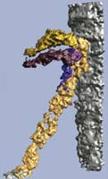
Hearing hinges on this nano-sized structure. Electron tomography was used to develop this surface rendering of a segmented tip link interconnecting two stereociliary membranes. The main tip link is shown in gold. Strands connecting the tip link and membrane are rendered in purple. The stereociliary membranes are portrayed in silver.
Their work opens the door for a more fundamental understanding of how hearing works. It may also lead to improved ways to treat some forms of hearing loss, which affects about ten percent of people.
The filaments help transform the mechanical vibrations of sound into electrical signals that can be interpreted by the brain. They are only four nanometers wide and 160 nanometers long (one nanometer is one-billionth of a meter), but if enough of them break, the world becomes silent. They're part of a sensory system that operates over a range of stimuli spanning six orders of magnitude. With it, people can hear a pin drop and a jet throttle to full power. No other sensory system in biology and the electrical engineering world is capable of this feat.
"It's one of the most beautifully deigned systems in the body," says Manfred Auer of Berkeley Lab's Life Sciences Division. "But how it really works remains a mystery. Our goal is to determine what the system looks like, so we can determine how it functions."
To do this, Auer and colleagues utilize electron tomography, which acquires hundreds of images of a structure at different angles, and reconstructs them into a three-dimensional composite. The technique yields highly detailed images of structures at the molecular scale.
No one had applied electron tomography to hearing research until about eight years ago, when Auer's team set out to learn more about one of the last unmapped components of the auditory system. The inner ear is lined with hair cells that sprout hair bundles. These hair bundles bob and sway in fluid — like a wheat field bending under the wind — as the ear drum absorbs sound waves.
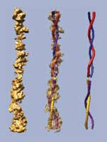
Zooming in even closer, each hair bundle is composed of individual hairs, also called stereocilia. Adjacent stereocilia are linked together by protein filaments, also known as tip links. When the stereocilia sway, the tip links stretch, which momentarily rips open a transduction channel that allows positively charged ions to stream into the hair cell. This initiates a neurotransmitter release that eventually reaches the nervous system. In this manner, a mechanical action — a channel prying open — is converted into an electrical signal and eventually something we hear as a chirp, beep, or voice.  These higher-magnification views of a tip link include a molecular model traced into the three-dimensional structure, revealing the link's twisting configuration.
"The system is incredible. But we still don't really know what constitutes the links, and we don't know how the hair bundle operates at the molecular level," says Auer.
That's beginning to change, thanks in part to Auer and colleagues' pioneering use of electron tomography to dissect the hair bundle at the molecular level. So far, they've reconstructed the hair-bundle links in three dimensions, and obtained highly accurate length measurements of the links, down to the molecular scale.
"One of the holy grails in structural cell biology is obtaining a molecular inventory of complex systems, and showing how the proteins work together to achieve their marvelous function," says Auer. "We're striving to develop such an inventory for the hair bundle."
Electron tomography studies of the hair bundle, in its cellular context, also enables the research team to decipher just how the hair bundle's capabilities are unmatched in nature and the man-made world. For example, how can it adapt to an extremely loud noise, and then quickly reconfigure itself to detect a whisper? And how can it be sensitive enough to detect the whisper, but not so sensitive that it detects every molecule colliding against the ear drum?
"If the system were any more sensitive, you would hear all of the molecules in the air bumping onto your ear drum, and go crazy," says Auer, adding that their recently obtained images are the first in a series of electron tomography explorations of hair cells.
"We know a good deal about how a hair bundle operates through clever electrophysiology experiments, but we need to know more, and for that we need to determine its molecular structure," says Auer. "Ultimately, we will get a molecular representation of this entire bundle, with all of its machinery, which will give us a fundamental insight into how the bundle works — and how hearing really works."
The research was funded by the National Institutes of Health, National Science Foundation, and the Department of Energy. It was reported in the June 9, 2008 issue of the Journal of the Association for Research in Otolaryngology.
Taken from www.lbl.gov/publicinfo.
Understanding Hearing, Molecule by Molecule
Share:

