Markers in our brain may help scientists to treat single-sided deafness.
A new discovery could help people suffering with single-sided deafness (SSD) find a treatment quicker - and could potentially lead to a cure.
SSD affects around 9,000 people per year in the UK, and around 60,000 per year in the US. It can be caused by a number of things - from viral infections to brain tumours - and is currently incurable and difficult to treat. Symptoms of SSD include impaired hearing, difficulty filtering out background noise, and difficulty determining sound direction.
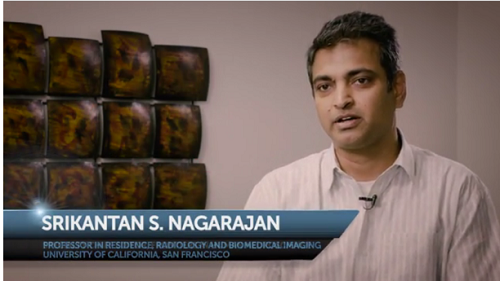
Srikantan S. Nagarajan is Professor in the Department of Radiology and Biomedical Imaging at UCSF, and a faculty member in the UCSF/UCB Joint Graduate Program in Bioengineering. In this video he discusses the latest research in brain plasticity. CREDIT: frontiersin.org (Click on image to launch video)
A major stumbling block to finding the best treatment has been the current lack of biomarkers against which to measure a treatment's efficacy, but Dr Srikantan Nagarajan and Dr Steven Cheung, a group of scientists based at the University of California have been looking at brain plasticity in response to the development of SSD. Their recent discovery could pave the way to the development of such biomarkers, and potentially, a cure.
Brain plasticity is the ability of the brain to modify its own structure and function in response to changes within the body (e.g., disease) or external factors. It is at the base of normal brain function: it helps us to learn and change our behaviour as children, and as adults can help us to overcome brain injuries, use prosthetic limbs, use hearing devices and more. With the development of new methods for studying brain plasticity, researchers are learning more about how our brains work and change, and how to harness this knowledge to our advantage.
This study used multi-modal imaging (MMI), one such new method that combines several brain-imaging techniques to look at changes in the brain. It is a powerful tool, helping scientists observe how the brain reorganises itself during learning and disease, and has many important applications. In this study, scientists used magnetoencephalographic imaging (MEGI), as well as fMRI scans, to look at the auditory cortices of 26 subjects - 13 people with SSD, and 13 with normal hearing.
There are two auditory cortices in the human brain; one in each hemisphere. The neurons in these cortices are arranged in a "tonotopic map", i.e., they are arranged according to the frequencies of sound to which they respond the best. So, neurons activated by one frequency of sound, are found near to neurons activated by a similar frequency.
The 26 subjects were exposed to sounds of different frequencies whilst the researchers monitored areas of activation in their auditory cortices. They observed the spread of neuron activation across the tonotopic maps in both brain hemispheres, and noticed significant differences between the two subject groups. The spread of activation was symmetrical across the cortices of the hemispheres of the brain in normal-hearing subjects, but not in SSD sufferers. In these cases, the spread of neuron activation was extended in one hemisphere and reduced in the other.
This discovery shows plasticity in both hemispheres of the brain in SSD sufferers, and is an important step towards developing biomarkers to help guide treatment choices. Ultimately, it may even be possible to use this plasticity to develop therapies to cure the condition: by using brain stimulation to restore a normal interhemispheric relationship, scientists may be able to restore normal auditory processing, returning sufferers to a life less affected by this hearing and communication handicap.
Source: https://www.eurekalert.org/pub_releases/2016-05/f-nbr051116.php


